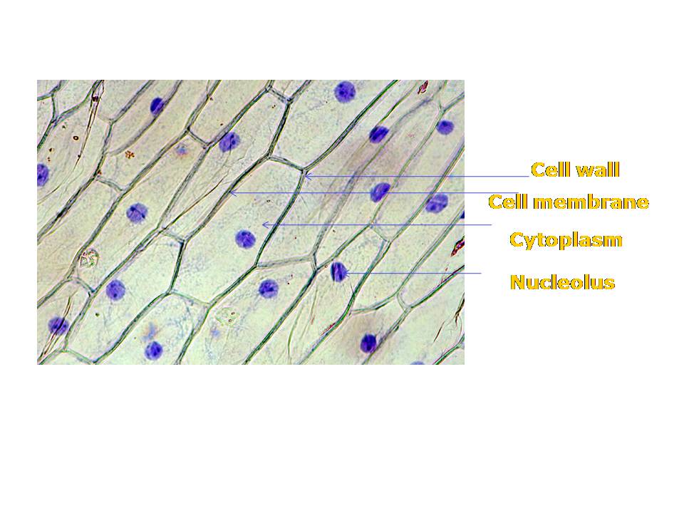
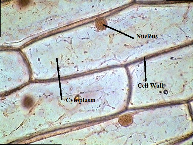
Labelled Diagram Of Onion Cell
Onion cell peel draw cytoplasm membrane vacuole showing figure brainly Labeled onion cell under microscope 40x Draw the figure of an onion peel showing cell
Onion Cells under Microscope - Saurabh Garg
Onion cell epidermal diagram labeled cells microscope under drawing skin epidermis lab bulb membrane mag observation preparation vacuole nucleus leaves Onion comentari deixa Onion cell stages under mitosis root tip microscope division different magnifications
Onion cell 400x lab microscope under labeled cells structure scoop science looked
Onion cell microscope under 40x micrograph labeled cells stock alamy microscopic section cepa allium rootOnion magnification 400x 100x Onion cells at 400x magnificationOnion labelled.
Onion cell epidermal peel sizeOnion epidermal cell labeled diagram Onion skin 200x plant slides dissectionOnion cell diagram drawing.

Onion_cells – biobiznews
The science scoop: onion cell labOnion cells under microscope Onion skin 200x « dissection connectionOnion cell stages of mitosis under microscope.
Onion cells microscope under magnified times cell 100x does genetics wallBiopedia: practicals .


Onion_Cells – BIOBIZNEWS

Onion Epidermal Cell Labeled Diagram - Wiring Diagram Pictures

draw the figure of an onion peel showing cell - Brainly.in

Onion Cell Stages Of Mitosis Under Microscope - Micropedia

Labeled Onion Cell Under Microscope 40x - Micropedia

Onion skin 200x « Dissection Connection

Onion Cell Diagram Drawing - lana1970

The Science Scoop: Onion Cell Lab

Onion Cells under Microscope - Saurabh Garg